

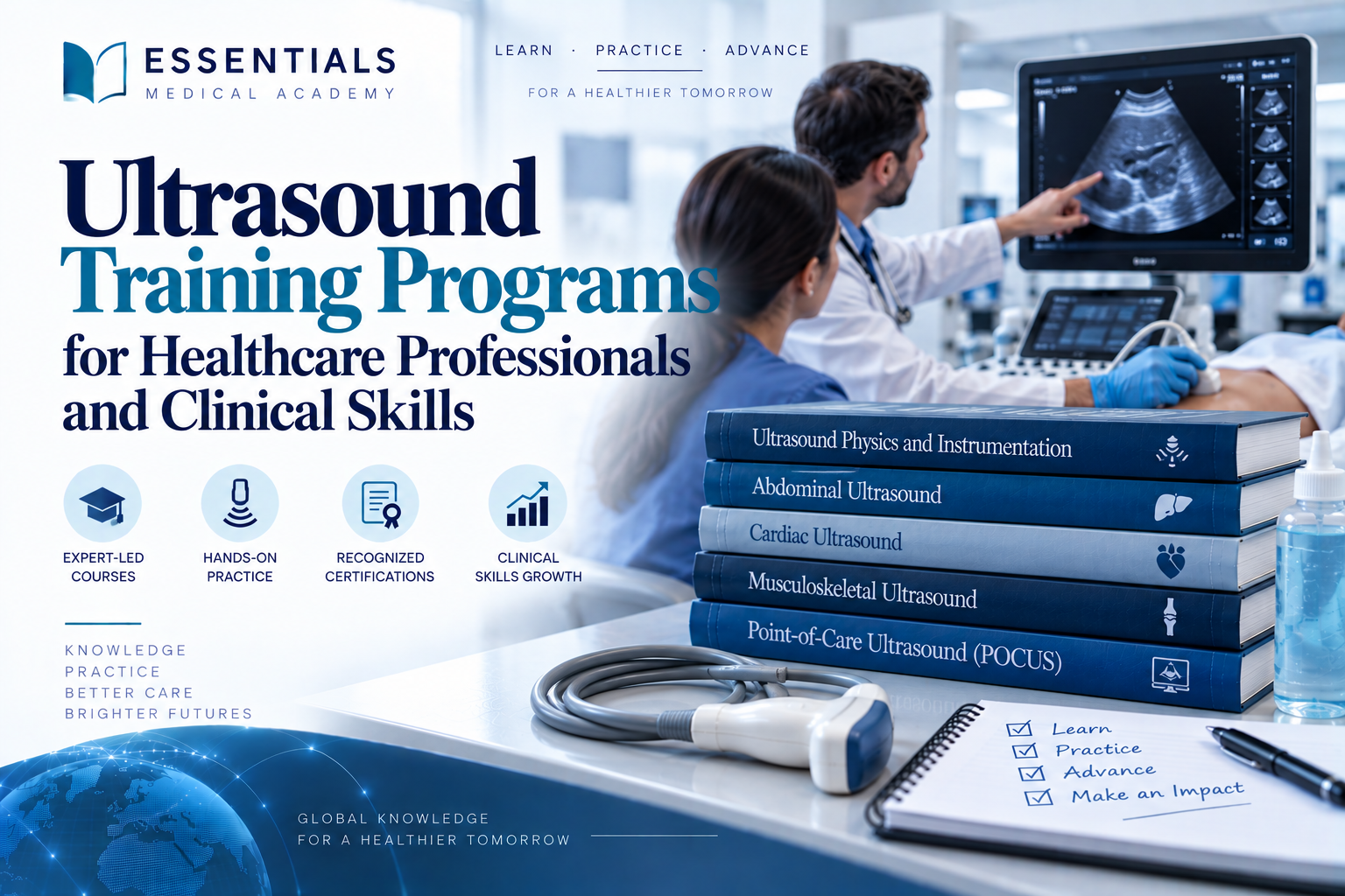
Ultrasound is a medical imaging technique that uses high-frequency sound waves to produce images of structures inside the human body. It allows healthcare professionals to examine organs, blood vessels, soft tissues, and developing pregnancies without using ionizing radiation. Because the procedure is generally noninvasive and can provide images in real time, it is widely used in hospitals, clinics, emergency departments, and specialized diagnostic facilities. Its versatility makes it valuable for evaluating many medical conditions and guiding selected clinical procedures.
The examination usually involves placing a small device called a transducer against the skin after applying a suitable gel. The transducer sends sound waves into the body and receives echoes reflected by internal structures. A computer processes these returning signals to generate images that help clinicians assess anatomy and identify abnormalities. Different tissues reflect sound differently, allowing the system to distinguish structures and display their appearance on a screen.
Medical applications include examining the liver, kidneys, gallbladder, thyroid gland, uterus, and other abdominal or pelvic organs. It can also help evaluate blood flow through vessels using Doppler techniques and support cardiac assessment through echocardiography. During pregnancy, imaging can provide information about fetal development and selected pregnancy-related concerns. In emergency settings, focused examinations may help clinicians rapidly investigate conditions such as internal bleeding or fluid accumulation.
The technology also supports image-guided procedures, including certain biopsies, fluid drainage techniques, and vascular access procedures. These applications require appropriate training, equipment, and clinical judgment to ensure that imaging findings are interpreted correctly and procedures are performed safely.
Although this imaging method offers many advantages, it has limitations. Image quality can be affected by body composition, bowel gas, the location of the target structure, and operator experience. Some conditions require additional investigations, such as computed tomography or magnetic resonance imaging. When used appropriately, this imaging approach supports timely assessment, informed clinical decisions, and patient-centered care across numerous medical specialties.
What Is Ultrasound Training for Healthcare Professionals?
Medical imaging education helps healthcare professionals understand how diagnostic equipment works, how images are acquired, and how findings contribute to clinical decision-making. A structured learning program can introduce the principles of image formation, patient preparation, equipment handling, anatomical recognition, and safe scanning practices. Depending on the learner’s background and professional goals, courses may focus on basic familiarity, a specific clinical application, or advanced skills requiring supervised practical experience and formal competency assessment.
Ultrasound training commonly covers the physics of sound waves, transducer selection, image optimization, scanning orientation, and recognition of normal anatomical structures. Learners explore how frequency, depth, gain, and probe position influence the resulting image. They may also study artifacts, common technical errors, infection prevention, patient positioning, and the importance of documenting findings accurately. Understanding these principles helps practitioners obtain useful images while recognizing when technical limitations may affect interpretation.
Practical instruction is particularly important because image acquisition requires coordination between visual interpretation, hand movements, and knowledge of anatomy. Supervised sessions allow participants to practice positioning the transducer, identifying anatomical landmarks, adjusting equipment settings, and obtaining standard views. Case-based discussions can demonstrate how imaging findings relate to symptoms, physical examination results, and other diagnostic information. Simulation may supplement hands-on teaching, although it cannot always replace supervised scanning on actual patients.
The appropriate course depends on professional responsibilities. Physicians may pursue point-of-care imaging or specialty-specific pathways, while nurses and other healthcare professionals may learn particular applications permitted within their roles. Formal credentials, supervised experience, and local regulations determine whether a practitioner can independently perform and interpret specific examinations. Completing a short course does not automatically authorize every imaging procedure.
When choosing a program, learners should review accreditation, faculty qualifications, curriculum, practical hours, assessment methods, and post-course supervision. Continuing education helps practitioners maintain image acquisition skills and remain aware of updated clinical guidance. A well-designed program combines theoretical knowledge, realistic practice, feedback, and professional accountability, enabling participants to use medical imaging appropriately and contribute to accurate, coordinated patient assessment.
Why Ultrasound Training Is Important in Modern Healthcare
Modern healthcare increasingly depends on timely diagnostic information to guide treatment, monitor disease, and evaluate patient responses. Medical imaging that can be performed quickly at the bedside offers practical advantages in emergency departments, inpatient wards, outpatient clinics, and specialized treatment settings. Developing relevant imaging competencies helps qualified professionals understand when an examination may answer a clinical question, how to obtain useful views, and when additional investigations or specialist consultation are necessary.
Ultrasound training is important because effective scanning involves more than operating a machine. Practitioners need to understand relevant anatomy, image formation, equipment settings, scanning techniques, and the limitations of the examination. Without sufficient knowledge, images may be incomplete or misleading, potentially affecting clinical decisions. Structured education helps learners recognize common artifacts, distinguish expected findings from possible abnormalities, and understand when an examination should be repeated or reviewed by an appropriately qualified specialist.
In emergency medicine, focused imaging may support the rapid assessment of selected conditions, including certain causes of abdominal pain, fluid accumulation, or cardiovascular instability. In other settings, it can assist with vascular access, evaluate musculoskeletal structures, monitor pregnancy, and support investigations of abdominal or pelvic symptoms. The specific applications depend on the clinician’s role, training, available equipment, and institutional protocols.
Appropriate education also promotes patient safety. Practitioners learn about infection prevention, correct transducer handling, patient identification, consent, documentation, and communication of important findings. They should understand that imaging results must be interpreted alongside the patient’s history, physical examination, and other relevant investigations. A normal scan does not necessarily exclude every serious condition, particularly when the examination is limited or the disease is difficult to visualize.
Competency development requires structured teaching, supervised practical experience, and reliable assessment. Continuing professional development helps clinicians maintain their skills and adapt to changes in equipment and clinical guidelines. Organizations should support appropriate training, clear scope-of-practice policies, and access to expert review when necessary. By combining technical proficiency with sound clinical judgment, healthcare professionals can use diagnostic imaging more effectively, support timely decisions, and improve coordination across patient care pathways.
How Ultrasound Technology Works in Medical Imaging
Medical imaging systems based on sound waves transform echoes from internal body structures into visual information. The process begins when an electronic system activates a transducer containing specialized elements that produce high-frequency sound pulses. These waves travel through body tissues and encounter boundaries between structures with different acoustic properties. Some energy is reflected toward the transducer, while the remainder continues deeper into the body. The returning echoes are detected and converted into electrical signals for processing.
A computer analyzes the timing and strength of these signals to estimate the location and appearance of internal structures. Echo timing helps determine depth, while signal intensity contributes to the brightness displayed in the resulting image. Repeated transmissions and rapid processing allow clinicians to view moving structures in real time. This capability is useful when examining the heart, observing fetal movement, evaluating organ motion, or guiding selected procedures.
Transducer frequency affects image resolution and penetration. Higher frequencies generally provide clearer detail in superficial tissues but do not penetrate as deeply. Lower frequencies are often used when examining deeper organs because they travel farther through tissue, although fine detail may be reduced. The operator also adjusts settings such as gain, depth, focus, and image orientation to optimize visualization for the examination being performed.
Doppler imaging uses changes in the frequency of returning echoes to assess the movement of blood. Depending on the technique, it can help estimate flow direction and velocity or display blood flow in selected vessels. Specialized approaches are used for cardiac examinations and other clinical applications requiring dynamic information.
Ultrasound examinations do not use ionizing radiation, which makes them suitable for many diagnostic purposes when performed appropriately. Nevertheless, image quality depends on equipment, patient factors, scanning technique, and operator expertise. Gas, bone, and certain tissue boundaries can limit visualization. Clinicians must therefore understand both the strengths and limitations of the technology before interpreting findings. Proper equipment maintenance, appropriate scanning protocols, and accurate clinical correlation help ensure that the images provide meaningful information for diagnosis and patient management.
The Role of Ultrasound in Clinical Diagnosis and Patient Care
Accurate diagnosis often requires combining a patient’s symptoms, medical history, physical examination, laboratory results, and appropriate imaging findings. Imaging based on sound waves can contribute valuable information by allowing clinicians to examine internal structures dynamically and, in many cases, directly at the bedside. Its accessibility and lack of ionizing radiation make it useful across numerous specialties, although the choice of examination must always reflect the clinical question and the patient’s circumstances.
Ultrasound supports the assessment of abdominal organs, including the liver, kidneys, gallbladder, and selected blood vessels. It may help investigate abdominal pain, identify certain fluid collections, and evaluate abnormalities that require further testing. Pelvic examinations can provide information about the uterus and ovaries, while obstetric applications help assess fetal development and selected pregnancy-related concerns. Cardiac imaging evaluates heart structure and function, and Doppler techniques provide information about blood movement through vessels.
In emergency and critical care settings, focused examinations may help clinicians assess selected causes of shock, investigate fluid accumulation, or evaluate certain respiratory and cardiac findings. Imaging can also guide procedures such as vascular access, selected injections, biopsies, and fluid drainage. When performed by appropriately trained professionals, visual guidance may improve targeting and help reduce avoidable procedural complications.
Ultrasound findings must be interpreted carefully. The accuracy of an examination depends on the clinical indication, image quality, equipment, operator experience, and the condition being investigated. Some abnormalities are difficult to detect, and a reassuring result does not always exclude significant disease. Clinicians may recommend additional imaging, laboratory tests, specialist assessment, or follow-up when findings remain uncertain or symptoms persist.
Patient communication is another essential part of diagnostic care. Professionals should explain the purpose of the examination, obtain appropriate consent, protect privacy, and communicate results through established clinical pathways. Accurate documentation supports continuity between healthcare teams and helps guide subsequent decisions. When used within its appropriate scope, this imaging method can improve clinical assessment, support treatment planning, and contribute to timely, individualized patient care without replacing comprehensive medical evaluation.
Who Should Enroll in Ultrasound Training Programs?
Medical education in diagnostic imaging can benefit healthcare professionals who want to improve their ability to assess patients and interpret clinical findings. Doctors, medical students, radiology residents, sonographers, and other qualified healthcare practitioners may find these learning opportunities valuable, depending on the course requirements and their professional responsibilities. Nurses and emergency care professionals may also benefit from introductory instruction when imaging skills are relevant to their clinical roles. Training typically covers image acquisition, basic anatomy, probe handling, image interpretation, and the appropriate use of diagnostic techniques in different clinical settings. Experienced practitioners can use further education to refresh their knowledge, explore advanced applications, and improve their confidence in patient assessment. Recent graduates may benefit from structured learning that connects theoretical concepts with supervised practical experience. Course selection should depend on the learner’s existing qualifications, intended specialty, and access to appropriate clinical supervision. Through Essentials, participants can strengthen their understanding of diagnostic imaging principles, develop relevant clinical competencies, and pursue continuing professional development. Combining theoretical instruction with hands-on practice helps learners build the skills necessary to support accurate assessments and safe, effective patient care within their authorized scope of practice.
Ultrasound Training for Emergency Medicine Professionals
Emergency medicine professionals need practical clinical skills to assess critically ill patients, identify urgent conditions, and make timely decisions in high-pressure environments. Training through Essentials can help healthcare providers develop their understanding of bedside imaging, recognize important anatomical structures, and evaluate findings that may influence immediate patient care. The curriculum may cover equipment handling, probe positioning, image acquisition, and the interpretation of common clinical presentations, including internal bleeding, cardiac abnormalities, and fluid accumulation. Hands-on practice, simulation exercises, and case-based discussions allow participants to connect theoretical knowledge with real-world scenarios. Learners also explore the limitations of diagnostic imaging, appropriate documentation, and situations that require additional investigations or specialist consultation. By combining structured education with supervised clinical practice, professionals can improve their confidence and contribute to more efficient patient assessment. Flexible learning opportunities enable doctors and other qualified healthcare providers to strengthen their expertise while balancing professional responsibilities. Continued practice and feedback from experienced instructors remain essential for maintaining competence and applying newly acquired skills safely in emergency settings.
Ultrasound Training for Doctors and Physicians
Medical professionals need strong diagnostic skills to evaluate symptoms, identify potential complications, and make informed decisions about patient care. Ultrasound training helps doctors and physicians develop practical knowledge of imaging principles and understand how real-time visualization can support clinical assessment. Depending on the course structure, participants may learn basic image acquisition, anatomical recognition, probe positioning, image interpretation, and appropriate documentation. These skills can complement physical examinations and help clinicians investigate specific clinical questions more efficiently.
Training programs may cover applications in emergency medicine, internal medicine, critical care, obstetrics, musculoskeletal assessment, and other specialties. Doctors can explore how imaging findings relate to symptoms, medical history, laboratory results, and examination findings. They also learn to recognize technical limitations, distinguish normal anatomical appearances from suspicious findings, and determine when additional investigations or specialist referrals are necessary. This approach encourages responsible clinical reasoning rather than reliance on images alone.
Practical sessions are particularly valuable because they allow participants to work with equipment under qualified supervision. Learners can practise scanning techniques, adjust image settings, identify anatomical landmarks, and receive feedback on their performance. Simulation exercises and supervised clinical practice may further strengthen confidence and consistency. The amount of hands-on experience varies between introductory courses and advanced specialty-specific programs.
When selecting a course, physicians should consider their specialty, previous experience, learning objectives, and the clinical settings in which they intend to use these skills. Accreditation, instructor qualifications, supervised practice, and competency assessment are also important considerations. Completing a course does not automatically grant independent practice privileges; clinicians must follow local regulations, professional standards, and institutional policies. Ongoing education helps maintain technical proficiency and supports safe, evidence-informed decisions that prioritize patient needs.
Ultrasound Training for Radiologists
Radiologists require a detailed understanding of diagnostic imaging, anatomical relationships, image quality, and the clinical significance of findings. Ultrasound training can support the development of advanced scanning and interpretation skills, helping specialists assess a broad range of conditions across different patient populations. Depending on their experience and subspecialty, participants may study abdominal imaging, pelvic examinations, vascular assessment, small-parts imaging, musculoskeletal applications, and image-guided procedures. Course content should align with the radiologist’s professional responsibilities and the requirements of their clinical practice.
A structured program may combine theoretical instruction with supervised scanning sessions, case reviews, and practical assessments. Participants can refine their understanding of acoustic principles, transducer selection, image optimization, Doppler techniques, and the factors that influence diagnostic accuracy. They may also learn systematic examination protocols, appropriate measurements, and consistent reporting methods. Reviewing representative cases helps learners connect imaging appearances with relevant clinical information while recognizing findings that require further investigation.
Quality assurance is another important component of professional development. Radiologists benefit from understanding equipment limitations, documenting technical difficulties, maintaining appropriate records, and applying standardized protocols. Training may also address patient communication, informed consent where applicable, infection prevention, and safe practice during image-guided interventions. These elements contribute to reliable examinations and support effective collaboration with referring clinicians.
When evaluating educational options, radiologists should consider faculty expertise, practical exposure, curriculum depth, accreditation, and opportunities for competency assessment. Advanced learners may benefit from specialty-focused workshops or supervised clinical placements rather than introductory material alone. However, course completion should not be interpreted as automatic authorization to perform every procedure or report every examination independently. Practice requirements differ by jurisdiction and institution. Continued professional education, peer review, and adherence to established guidelines help radiologists maintain high standards and provide imaging services that support appropriate patient management.
Ultrasound Training for Sonographers and Radiologic Technologists
Sonographers and radiologic technologists play an important role in producing diagnostic images and supporting accurate clinical evaluations. Ultrasound training helps these professionals strengthen their understanding of equipment operation, scanning protocols, patient positioning, anatomical landmarks, and image optimization. Depending on their existing qualifications, learners may focus on general imaging, abdominal examinations, vascular studies, obstetric and gynecological applications, or other specialty areas. The appropriate curriculum depends on local professional requirements, workplace responsibilities, and previous clinical experience.
Practical instruction is central to developing consistent scanning techniques. Participants can learn how to select suitable transducers, adjust gain and depth, optimize focus, recognize artifacts, and obtain images that answer the examination request. Supervised practice also provides opportunities to improve hand-eye coordination, adapt techniques to different body types, and manage examinations when patient mobility or positioning is limited. Case-based learning helps connect technical decisions with the diagnostic purpose of each procedure.
Patient safety and professional communication are equally important. Technologists should understand how to explain examinations clearly, protect privacy, follow infection-prevention procedures, and respond appropriately when patients experience discomfort or anxiety. Training can also reinforce accurate documentation, correct patient identification, equipment care, and timely communication of urgent concerns through established clinical pathways. Recognizing the limits of one’s role is essential, particularly when unexpected or potentially significant findings arise.
Before enrolling, learners should review course prerequisites, practical hours, instructor qualifications, assessment methods, and recognition by relevant professional bodies. Introductory programs may be suitable for learners developing foundational knowledge, while experienced practitioners may prefer advanced modules or specialty-specific education. Completing a short course does not necessarily replace an accredited qualification or satisfy licensing requirements. Regular practice, continuing education, and feedback from qualified supervisors help professionals maintain technical competence and contribute effectively to multidisciplinary patient care.
Ultrasound Training for Nurses
Nurses work across diverse clinical environments, including emergency departments, intensive care units, inpatient wards, outpatient clinics, and community healthcare services. Ultrasound training can help eligible nursing professionals understand how bedside imaging may contribute to focused assessment, clinical monitoring, and communication with other members of the healthcare team. The appropriate course depends on the nurse’s role, prior experience, institutional policies, and the specific applications permitted within their professional scope of practice.
Educational programs may introduce basic imaging principles, equipment handling, probe orientation, anatomical recognition, and focused assessment protocols. Depending on the curriculum, nurses may explore applications relevant to vascular access, bladder assessment, selected emergency evaluations, or other approved bedside procedures. Supervised demonstrations and practical exercises help learners understand how to position the probe, optimize images, identify limitations, and recognize when findings are unclear. Training should emphasize that imaging complements clinical assessment rather than replacing a complete evaluation or the judgment of appropriately qualified practitioners.
Patient-centered practice remains essential throughout the learning process. Nurses should communicate the purpose of an examination, maintain privacy, obtain consent when required, and follow infection-control procedures. They also need to document observations according to workplace protocols and escalate concerning findings through established clinical pathways. Understanding when to seek assistance is particularly important because image quality and interpretation can be affected by patient factors, equipment settings, and the operator’s experience.
Before choosing a program, nurses should verify its suitability for their specialty and whether it is recognized by the relevant employer or professional authority. Useful features include qualified instructors, supervised hands-on practice, clear learning outcomes, competency assessment, and guidance on documentation. Course completion alone does not authorize every nursing professional to perform or interpret all imaging examinations independently. Ongoing education and regular supervised practice can help nurses develop appropriate skills while maintaining professional accountability and prioritizing patient safety.
Ultrasound Training for Critical Care and ICU Professionals
Critical care professionals manage patients who may experience rapid changes in their clinical condition, requiring timely assessment and coordinated treatment decisions. Ultrasound training can help eligible clinicians develop practical skills for focused bedside evaluation and improve their understanding of how imaging findings relate to a patient’s overall condition. Depending on the curriculum, training may introduce lung assessment, focused cardiac evaluation, abdominal imaging, vascular access guidance, and examinations that support the assessment of fluid status. These applications can be particularly relevant when transporting an unstable patient to another department presents additional challenges.
A structured educational program typically combines theoretical learning with supervised practical sessions. Participants may study basic imaging principles, probe selection, image optimization, anatomical landmarks, and systematic scanning protocols. Case-based exercises can help learners understand how findings contribute to clinical reasoning and when further diagnostic investigations are appropriate. Training should also address common artifacts, technical limitations, and situations in which an examination cannot provide a reliable answer.
For intensive care teams, effective communication is an essential part of the learning process. Clinicians must document their observations appropriately, communicate significant concerns, and collaborate with radiologists, cardiologists, and other specialists when necessary. Clear protocols help ensure that bedside findings are interpreted within the context of symptoms, physical examination results, laboratory tests, and the patient’s medical history.
When selecting a course, professionals should consider instructor qualifications, supervised scanning opportunities, assessment methods, and relevance to their clinical responsibilities. Introductory workshops may provide foundational knowledge, while advanced programs can focus on specialty-specific applications. Completing a course does not automatically establish independent competency or expand a clinician’s authorized scope of practice. Local regulations and institutional policies remain essential. Continued practice, feedback, and periodic competency assessment help critical care professionals maintain appropriate skills and use imaging responsibly to support patient management.
Ultrasound Training for Obstetricians and Gynecologists
Obstetricians and gynecologists use diagnostic imaging to support the evaluation of pregnancy, reproductive health, and various gynecological conditions. Ultrasound training can help these specialists strengthen their understanding of pelvic anatomy, examination techniques, image interpretation, and the clinical significance of findings. Depending on the course level, educational content may include early pregnancy assessment, fetal development, placental location, amniotic fluid evaluation, uterine abnormalities, ovarian findings, and the use of Doppler techniques where appropriate.
A comprehensive program combines theoretical instruction with supervised practical experience. Participants may learn how to select suitable transducers, obtain standard views, optimize image quality, and recognize anatomical landmarks. Training can also address differences between transabdominal and transvaginal approaches, including their indications, limitations, and relevant consent considerations. Case discussions allow specialists to compare imaging findings with clinical history and examination results while identifying situations that require additional testing or specialist referral.
Professional development in this field also emphasizes patient communication and responsible interpretation. Clinicians should explain the purpose and limitations of examinations, protect confidentiality, document findings accurately, and communicate important concerns through appropriate clinical pathways. In pregnancy care, understanding the limits of imaging is particularly important because not every fetal condition can be detected, and examination results must be interpreted within the appropriate clinical context.
When choosing a program, obstetricians and gynecologists should assess the curriculum, faculty experience, supervised practice hours, competency evaluations, and recognition by relevant professional organizations. Beginners may benefit from foundational courses, while experienced specialists may prefer advanced modules in specific clinical applications. Course attendance alone does not guarantee proficiency in every examination or procedure. Ongoing education, regular practice, and adherence to professional guidelines help clinicians maintain their skills and provide safe, evidence-informed reproductive and maternity care.
Ultrasound Training for Cardiologists
Cardiologists evaluate cardiovascular conditions that may affect the heart, blood vessels, and overall circulation. Ultrasound training can support their understanding of cardiac imaging principles and help them develop skills relevant to assessing cardiac structure, function, and blood flow. Depending on the course level, participants may study echocardiographic views, ventricular function, valve abnormalities, Doppler measurements, and selected applications in acute cardiac assessment. The curriculum should match the learner’s previous experience and intended clinical responsibilities.
A well-designed program combines foundational theory, image demonstrations, supervised scanning, and case-based interpretation. Learners may practise identifying cardiac chambers, recognizing standard imaging windows, adjusting equipment settings, and understanding how image quality affects diagnostic confidence. Instruction can also cover the limitations of particular views, the significance of common artifacts, and the importance of integrating imaging results with symptoms, examination findings, electrocardiograms, and laboratory investigations.
Advanced education may explore more specialized techniques, including assessment of complex valve disease, evaluation of ventricular performance, and selected applications involving hemodynamic measurements. These topics require appropriate prerequisites and supervised experience. Training should emphasize standardized protocols, accurate reporting, quality assurance, and recognition of findings that warrant urgent review or additional investigation. Collaboration with other cardiovascular specialists can further strengthen clinical decision-making and continuity of care.
Cardiologists selecting a course should consider instructor expertise, accreditation, practical scanning opportunities, case diversity, and formal competency assessment. Introductory courses may be appropriate for clinicians developing basic familiarity, whereas advanced practitioners may need structured specialty training. A certificate of attendance does not automatically qualify a participant to perform or interpret every cardiac examination independently. Local licensing rules, institutional requirements, and professional standards determine the scope of authorized practice. Continuing education, supervised experience, and peer review help cardiologists maintain proficiency and use imaging findings appropriately when planning patient care.
Ultrasound Training for Anesthesia Professionals
Anesthesia professionals work in operating rooms, procedural areas, recovery units, and other settings where accurate assessment and safe procedural techniques are essential. Ultrasound training can help anesthesiologists and other appropriately qualified practitioners develop skills that support selected clinical assessments and image-guided interventions. Depending on the program, applications may include peripheral nerve blocks, vascular access, assessment of relevant anatomical structures, and focused cardiovascular evaluation in suitable clinical circumstances.
A structured course typically introduces imaging principles, transducer selection, probe orientation, image optimization, and recognition of important anatomical landmarks. Practical sessions allow participants to observe demonstrations, practise scanning techniques, and receive feedback from experienced instructors. For regional anesthesia, learners may study the relationship between nerves, surrounding tissues, and nearby vessels, while also reviewing approaches to needle visualization and the recognition of structures that should be avoided. Training should reinforce safe procedural planning rather than relying on image recognition alone.
Patient safety remains central throughout the learning process. Professionals should understand relevant contraindications, infection-prevention procedures, consent requirements, monitoring standards, and strategies for responding to complications. They must also recognize technical limitations and know when to request assistance or use an alternative approach. Simulation-based practice can help learners develop coordination and procedural familiarity before performing techniques in supervised clinical environments.
When choosing an educational program, anesthesia professionals should review the curriculum, instructor qualifications, hands-on practice hours, assessment methods, and relevance to their workplace responsibilities. Introductory courses can establish foundational knowledge, while advanced workshops may address particular blocks or specialized assessment techniques. Completing a course does not automatically establish independent procedural competence or confer additional professional privileges. Local regulations and institutional protocols remain decisive. Ongoing supervised practice, competency reviews, and continuing education help practitioners maintain appropriate skills and support safe, individualized anesthesia care.
Ultrasound Training for Allied Health Professionals
Allied health professionals can strengthen their clinical knowledge and expand their practical abilities through specialized education designed to support patient assessment and collaborative healthcare delivery. Training through Essentials introduces learners to the fundamentals of medical imaging, including equipment operation, patient positioning, image acquisition, and basic interpretation principles relevant to their professional responsibilities. Participants can explore how bedside imaging supports clinical examinations, helps identify potential abnormalities, and contributes to informed decisions alongside other diagnostic methods. The curriculum may include anatomical identification, infection prevention, patient communication, documentation, and recognition of findings that require referral to qualified specialists. Practical demonstrations and case-based learning help participants connect theoretical concepts with clinical situations while understanding the importance of accuracy and patient safety. Ultrasound education can be particularly valuable for eligible professionals whose roles involve diagnostic support, rehabilitation, or specialized clinical assessments, depending on their qualifications and local regulations. Flexible online learning allows healthcare workers to develop their knowledge around existing responsibilities, while supervised practice helps reinforce essential techniques. By pursuing continued professional development, allied health professionals can improve interdisciplinary communication, strengthen their confidence, and contribute more effectively to coordinated, patient-centered care.
Ultrasound Training for Medical Students and Recent Graduates
Medical students and recent graduates need a strong foundation in clinical assessment, anatomy, and diagnostic reasoning as they prepare for professional practice. Ultrasound training can introduce learners to practical imaging concepts and demonstrate how bedside visualization may complement history-taking and physical examination. Early exposure helps students understand the relationship between anatomical structures and clinical findings while developing familiarity with equipment, transducer positioning, and basic image acquisition techniques.
A suitable introductory program typically combines short theoretical lessons, demonstrations, supervised scanning, and case-based discussions. Students may learn to recognize common anatomical landmarks, optimize image quality, identify basic imaging artifacts, and understand the limitations of different examination approaches. Depending on the curriculum, learning activities may cover abdominal assessment, focused cardiac views, lung examination, vascular access guidance, and selected musculoskeletal applications. These sessions should be appropriate for the learner’s level and should not replace formal instruction in clinical assessment.
Practical experience is particularly valuable because it allows learners to connect classroom knowledge with real clinical scenarios. Under qualified supervision, students can practise probe movements, identify structures in different imaging planes, and receive feedback on image acquisition. Case discussions can also help them understand why a finding must be interpreted alongside symptoms, medical history, physical examination results, and other investigations.
When selecting a course, students should consider the instructor’s qualifications, the amount of hands-on practice, the availability of feedback, and the relevance of the curriculum to their future specialty. Recent graduates may benefit from additional supervised sessions that reinforce foundational skills before they enter more demanding clinical environments. Completing a short course does not automatically authorize independent scanning or interpretation. Professional regulations, institutional policies, and competency requirements still apply. Continued learning and supervised clinical experience help new healthcare professionals build confidence responsibly while maintaining patient safety and appropriate professional boundaries.
Ultrasound Training for Experienced Healthcare Professionals
Experienced healthcare professionals often need continuing education to maintain their clinical skills, adapt to evolving technologies, and address the demands of their specialties. Ultrasound training can provide opportunities to refine existing techniques, explore advanced applications, and improve understanding of how imaging findings contribute to patient assessment. The most useful program is one that matches the participant’s professional background, current competencies, and intended clinical responsibilities rather than repeating material already mastered.
Advanced courses may focus on specialty-specific examinations, image optimization, interpretation of complex findings, procedural guidance, or structured clinical assessment. Depending on the learner’s role, topics may include focused cardiac imaging, vascular evaluation, musculoskeletal applications, emergency assessment, and selected image-guided interventions. Case-based teaching can help experienced practitioners recognize diagnostic uncertainty, understand technical limitations, and decide when further investigation or specialist consultation is appropriate.
Hands-on practice remains essential, even for clinicians who regularly use imaging equipment. Supervised sessions allow participants to review scanning protocols, refine probe manipulation, compare image quality, and receive feedback on technique. Peer discussions and clinical case reviews can also reveal differences in practice and reinforce consistent documentation. Programs that include formal assessment may help learners identify gaps in knowledge and establish clear objectives for continued professional development.
Before enrolling, professionals should examine course prerequisites, instructor expertise, practical hours, assessment standards, and recognition by relevant professional bodies. Advanced workshops may be more valuable than introductory courses for participants with established experience. However, a certificate alone does not guarantee independent competency, expand a professional’s authorized scope, or replace required credentials. Local regulations and institutional policies determine which examinations and procedures a clinician may perform. Regular practice, continuing education, and quality review help experienced professionals maintain appropriate standards and integrate imaging into clinical decisions responsibly.
Core Competencies Required in Medical Ultrasound
Healthcare professionals who use diagnostic imaging need a combination of technical knowledge, anatomical understanding, clinical reasoning, and professional responsibility. Ultrasound competency involves more than operating equipment or obtaining recognizable images. Practitioners must understand the purpose of each examination, select suitable scanning techniques, evaluate image quality, and recognize when technical limitations prevent a reliable conclusion. These abilities support consistent examinations and help reduce the risk of misinterpretation.
Foundational knowledge includes acoustic principles, transducer types, image orientation, common artifacts, and the effects of settings such as depth, gain, and focus. Professionals also need a sound understanding of the anatomy relevant to their clinical specialty. Practical competencies include positioning the patient appropriately, obtaining standard views, identifying anatomical landmarks, and following established scanning protocols. The required level of proficiency varies according to the examination, professional role, and local practice requirements.
Clinical reasoning is another essential component. Practitioners should connect imaging observations with the patient’s symptoms, medical history, physical examination, and other available evidence. They must distinguish between a technically inadequate study, an uncertain finding, and an observation that requires further assessment. Recognizing the limits of an examination and seeking specialist input when necessary are important indicators of safe practice.
Professional competencies extend to patient communication, informed consent where applicable, privacy, infection prevention, accurate documentation, and appropriate escalation of concerning findings. Practitioners should also understand equipment maintenance, quality assurance, and the importance of continuing education. Competency assessments may combine theoretical examinations, observed practical tasks, case interpretation, and supervised clinical experience. Course attendance alone is not sufficient evidence of independent proficiency. Regular feedback, peer review, and adherence to professional guidelines help clinicians maintain their skills while ensuring that imaging contributes appropriately to patient care.
Essential Clinical Skills for Ultrasound Professionals
Professionals who perform diagnostic imaging need practical abilities that support accurate examinations, effective communication, and safe patient care. Ultrasound practice requires coordinated use of technical skills and clinical judgment, beginning with a clear understanding of the examination request. Before scanning, practitioners should verify patient identity, review relevant clinical information, explain the procedure, and follow applicable consent and privacy requirements. Appropriate preparation helps ensure that the examination addresses the clinical question and respects patient needs.
Technical skills include selecting the correct transducer, positioning the patient, orienting the probe, adjusting image settings, and obtaining appropriate views. Practitioners must recognize anatomical landmarks and understand how movement, pressure, body habitus, and equipment settings can influence image quality. They should also identify common artifacts and recognize when an examination is incomplete or technically limited. Following a systematic protocol can improve consistency and reduce the likelihood of overlooking relevant structures.
Interpretive and communication skills are equally important. Professionals need to describe observations accurately, document technical limitations, and avoid making conclusions beyond their training or authorized role. Unexpected findings should be handled according to established clinical pathways, including timely communication with the appropriate clinician. Effective collaboration with radiologists, physicians, nurses, and other team members helps integrate imaging information into the broader care plan.
Patient safety skills include infection prevention, equipment hygiene, appropriate handling of transducers, and awareness of relevant examination precautions. Professionals should also understand when a different diagnostic method or specialist assessment may be more suitable. Practical workshops, simulation, supervised clinical experience, and structured feedback can help learners develop these abilities progressively. Competency should be assessed according to the specific tasks practitioners are expected to perform. Ongoing education, regular review of clinical practice, and adherence to professional standards help maintain technical reliability and support responsible use of imaging in diverse healthcare settings.
